Causes
It is unsure what causes lipomas to grow. They are inherited (passed down through families). You're more likely to develop a lipoma if someone in your family has one.
Some conditions cause multiple lipomas to form on the body. Lipoma-causing conditions include:
- Dercum's disease: This rare disorder causes painful lipomas to grow, most often on the arms, legs and trunk. It's also called adiposis dolorosa or Anders' syndrome.
- Gardner syndrome: A form of a disorder called familial adenomatous polyposis (FAP), Gardner syndrome causes lipomas and a range of health problems.
- Hereditary multiple lipomatosis: Also called familial multiple lipomatosis, this disorder is inherited (passed down through families).
- Madelung's disease: This condition occurs most often in men who drink alcohol excessively. Also called multiple symmetric lipomatosis, Madelung's disease causes lipomas to grow around the neck and shoulders.
Diagnosis
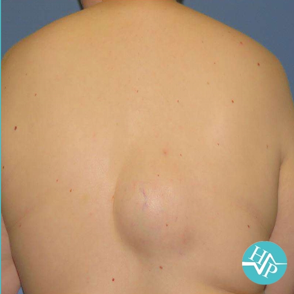
Providers usually diagnose a lipoma during a physical examination. Your provider will touch the lipoma and ask if it's painful or tender. You may need a biopsy to confirm that the lipoma isn't cancer. During this procedure, your provider removes a sample of the lipoma and sends it to a lab for testing.
Oftentimes, these may be mistaken for a cyst. To see a clear picture of this lump, your provider may order an imaging test such as an ultrasound, magnetic resonance imaging (MRI) scan, or computed tomography (CT) scan. These imaging studies help your provider determine if it is a lipoma versus a cyst. It can also help identify the lipoma's location and how deep it is if it has blood vessels and whether it's pressing against nerves or other tissues.
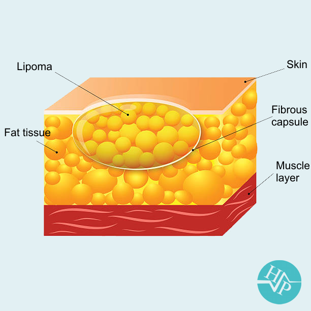
Types of lipomas
All lipomas are made of fat. Some lipomas also contain blood vessels or other tissues. There are several types of lipomas, including:
Angiolipoma: This type contains fat and blood vessels. Angiolipomas are often painful.
Conventional: The most common type, a conventional lipoma contains white fat cells. White fat cells store energy.
Fibrolipoma: Fat and fibrous tissue make up this type of lipoma.
Hibernoma: This kind of lipoma contains brown fat. Most other lipomas contain white fat. Brown fat cells generate heat and help regulate body temperature.
Myelolipoma: These lipomas contain fat and tissues that produce blood cells.
Spindle cell: The fat cells in these lipomas are longer than they are wide.
Pleomorphic: These lipomas have fat cells of various sizes and shapes.
Treatment
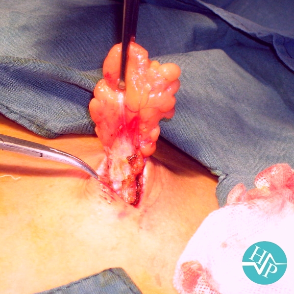
Larger lipomas are best removed through incisions made in the skin overlying the lipoma. The incisions are configured like a fusiform excision following the skin tension lines and are smaller than the underlying tumor. The central island of skin to be excised is grasped with a hemostat, or Allis clamp, which is used to provide traction for the removal of the tumor. Dissection is then performed beneath the subcutaneous fat to the tumor. Any tissue cutting is performed under direct visualization using a no. 15 scalpel or scissors around the lipoma. Care must be taken to avoid nerves or blood vessels that may lie just beneath the tumor.
Once a portion of lipoma has been dissected from the surrounding tissue, hemostats or clamps can be attached to the tumor to provide traction for removal of the remainder of the growth. Once it is freed, the lipoma is delivered as a whole. The surrounding tissue in the hole can be palpated to ensure complete removal of the tumor.
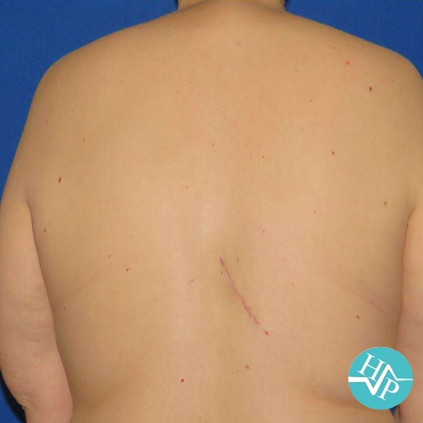
Adequate hemostasis is achieved following the removal of the lipoma using hemostats or suture ligation. The dead space is closed beneath the skin using buried, interrupted 3-0 or 4-0 Vicryl sutures. Occasionally drains may have to be placed to prevent fluid accumulation, but this should be avoided if possible. The skin is then closed with interrupted 4-0 or 5-0 nylon sutures. A pressure dressing is placed to reduce the incidence of hematoma formation. The patient is given routine wound care instructions, and the wound is checked in two to seven days. The sutures are removed after seven to
21 days, depending on the body location. Specimens should be submitted for histologic analysis.